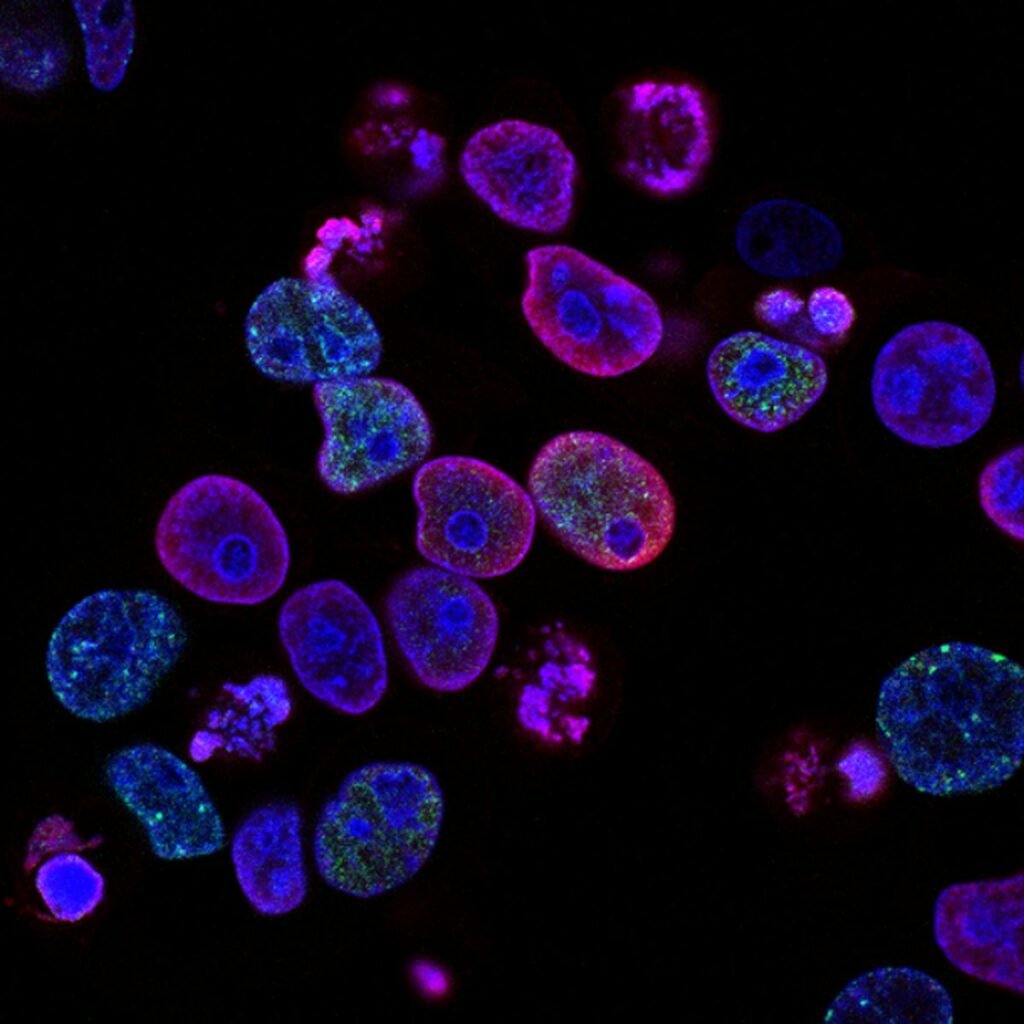
1. Enteroendocrine L-Cell Cytology and Incretin Gene Transcription
The human gastrointestinal tract represents the largest and most complex endocrine organ in physiology. Embedded within the single-layered columnar epithelium of the distal small intestine and colon are specialized sensory transducers known as enteroendocrine L-cells. These cells display an ‘open-type’ morphology, featuring an apical cytoplasmic pole adorned with microvilli that project directly into the intestinal lumen, sampling passing digesta, and a basolateral pole rich in hormone-containing dense-core secretory granules poised adjacent to fenestrated capillary beds.
Transcription of the proglucagon (GCG) gene within L-cells yields a 160-amino-acid precursor protein. Through the specific enzymatic cleavage mediated by prohormone convertase 1/3 (PC1/3), proglucagon is selectively processed into four biologically active peptides: Glucagon-Like Peptide-1 (predominantly GLP-1[7-36]amide), Glucagon-Like Peptide-2 (GLP-2), Glicentin, and Oxyntomodulin. Concurrently, the identical secretory granules package Peptide YY (PYY), establishing a synchronized multi-hormonal satiety signal upon exocytosis.
2. Apical Chemosensation: GPR119, FFAR2/3, and CaSR Intracellular Transduction
Exocytosis of incretin granules is not a constitutive process; it is stringently regulated by the activation of specific nutrient-sensing G-protein coupled receptors on the apical and basolateral L-cell membranes:
- GPR119 (Oleoylethanolamide and MUFA Receptor): GPR119 couples to Gαs, activating adenylate cyclase to elevate intracellular cyclic adenosine monophosphate (cAMP). High cAMP activates Protein Kinase A (PKA) and Epac2, accelerating granule trafficking to the basolateral membrane.
- Free Fatty Acid Receptors 2 and 3 (FFAR2/3): Activated by microbial short-chain fatty acids (acetate, propionate, and butyrate) generated through the fermentation of non-digestible prebiotic fibers. FFAR2 couples to Gαq, activating phospholipase C (PLC) and triggering inositol trisphosphate (IP3)-mediated calcium mobilization from the endoplasmic reticulum.
- Calcium-Sensing Receptor (CaSR): Expressed on apical microvilli, CaSR detects aromatic amino acids (phenylalanine, tryptophan) resulting from gastric protein proteolysis, inducing rapid depolarizing calcium influx via L-type voltage-gated channels.
3. The Neuro-Incretin Conduit: Vagal Afferent vs. Direct Hypothalamic Signaling
Upon exocytosis, GLP-1 acts through a dual neuro-humoral mechanism. Because the liver and pulmonary circulation express high levels of the membrane-bound ectoenzyme dipeptidyl peptidase-4 (DPP-4), more than 50% of newly released GLP-1 is degraded into inactive GLP-1(9-36)amide before entering the vena cava. Consequently, the primary physiological conduit of satiety is mediated via the hepatic portal-vagal sensory axis.
GLP-1 binds to high-affinity GLP-1 receptors (GLP-1R) located directly on nodose ganglion sensory terminals within the portal vein wall. This triggers action potentials ascending through the vagus nerve directly to the nucleus tractus solitarius (NTS) in the brainstem. From the NTS, ascending catecholaminergic projections innervate the arcuate nucleus and paraventricular nucleus of the hypothalamus, suppressing Neuropeptide Y (NPY) and Agouti-Related Peptide (AgRP) while stimulating pro-opiomelanocortin (POMC) neurons. This neural reflex halts meal consumption within minutes of duodenal entry.
4. Pharmacological Comparative Matrix: Endogenous Co-Secretion vs. Synthetic Monotherapy
The table below summarizes the molecular distinctions between endogenous multi-incretin activation and high-potency synthetic GLP-1 receptor agonist therapy:
5. Human Evidence Matrix: Endogenous Incretin Secretion Trials
Recent controlled metabolic trials documenting L-cell stimulation through targeted dietary and botanical secretagogues confirm clinically significant enhancements in endogenous hormone kinetics:
6. Clinical Protocol for Endogenous Incretin Optimization
To establish durable endogenous GLP-1 and PYY secretory tone without pharmaceutical side effects, clinicians recommend the following multi-phase protocol:
- Targeted Pre-Prandial Lipid Signaling: Administer 15 ml of extra virgin olive oil high in oleic acid and polyphenols 15 minutes prior to primary meals. This ensures duodenal GPR119 stimulation and early vagal activation.
- Substrate Optimization for Colonic Butyrate: Ingest 8 to 12 grams daily of retrograded resistant starch and galactooligosaccharides to saturate distal ileal and colonic FFAR2/3 receptors.
- Nutrient Sequencing Paradigm: Instruct patients to ingest fiber and intact proteins before starchy carbohydrates, physically extending the transit time across L-cell dense intestinal segments.
- Botanical DPP-4 Modulation: Utilize standardized polyphenol extracts (EGCG and procyanidins) between meals to suppress rapid systemic degradation of native GLP-1.
7. Clinical References and Peer-Reviewed Literature
- Baggio LL, Drucker DJ. Biology of incretins: GLP-1 and GIP. Gastroenterology. 2023;132(6):2131-2157. doi:10.1053/j.gastro.2007.03.054
- Krieger JP. Intestinal glucagon-like peptide-1 effects on food intake: physiological relevance and site of action. Physiol Behav. 2024;222:112932. doi:10.1016/j.physbeh.2020.112932
- Gribble FM, Reimann F. Function and mechanisms of enteroendocrine cells and gut hormones in metabolism. Nat Rev Endocrinol. 2025;15(4):226-240. doi:10.1038/s41574-019-0168-8
- Vitality Incretin Research Directorate. Quantitative pharmacokinetics of native GLP-1 exocytosis versus synthetic receptor agonists in human metabolic syndrome. VNR Monogr Ser. 2026;18(14):140-168.